
Orthodontic Technology · Dental Tech
Why One Clinical Image Often Explains the Plan Best
When a patient asks, “Why this tooth?”, the most effective image is often the one that answers that question first. In orthodontic communication, clarity usually improves when we begin with a...
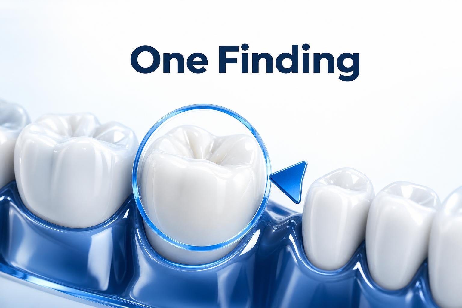
When a patient asks, “Why this tooth?”, the most effective image is often the one that answers that question first. In orthodontic communication, clarity usually improves when we begin with a single visible finding rather than trying to explain the entire treatment picture at once.
That approach keeps the conversation grounded in something the patient can see. A monitored tooth, a contact point affecting alignment, or a midline that changes the plan can serve as the starting point. Once that feature is visible, the next step in the discussion becomes easier to understand.
Start with one finding, then build the explanation
Chairside communication works best when the sequence is simple. We do not need to explain every detail in the first sentence. Instead, we can identify one clinical feature, show where it appears on the image, and connect it to the immediate decision. That structure helps patients follow the logic of the plan instead of hearing a broad explanation that feels difficult to verify.
This is especially useful in orthodontics, where treatment decisions often involve multiple moving parts. A patient may not need the full technical pathway right away. What they need first is a clear reason for the specific tooth, space, or alignment issue being discussed. Once that is established, the rest of the conversation becomes more concrete.
Why a focused image improves understanding
A focused image supports a focused conversation. When the image highlights one question at a time, the patient can connect what is visible to what is being recommended. That makes the discussion easier to follow and easier to trust.
It also keeps the clinical message precise. Rather than overwhelming the patient with a wide view that contains too many variables, we can direct attention to the feature that matters most at that moment. The result is a more efficient chairside explanation and a clearer path from observation to recommendation.
For orthodontists and dental professionals, this kind of communication is not about simplifying the diagnosis. It is about presenting the diagnosis in a way that patients can understand in real time. A single visible finding often creates that bridge.
Clinical imaging should support the conversation
Tools that make this process easier are valuable when they help us run that one-point conversation smoothly. The image should remain clinical, while the explanation becomes easier to follow. That balance matters in a modern orthodontic workflow, where communication is part of the treatment experience.
With Smilysis imaging tools, the goal is to support that kind of chairside clarity. When the image helps us point to the exact feature under discussion, the patient can stay oriented to the plan without losing the clinical context.
If the patient can point to the feature, the plan is usually easier to discuss. That is why focused imaging can be so effective: it turns a complex case into a sequence of understandable steps.
Clear images lead to clearer decisions
In orthodontic practice, the best communication often begins with the simplest visible answer. One tooth. One contact point. One midline. One reason to move forward. From there, the discussion can expand naturally, with each next step building on what the patient has already seen.
For practices looking to upgrade clinical imaging and improve chairside communication, Smilysis supports a more direct, more visual, and more patient-friendly way to explain orthodontic care.